Caries treatment under a microscope
Learn more
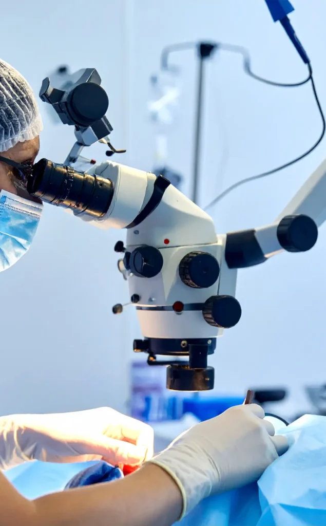
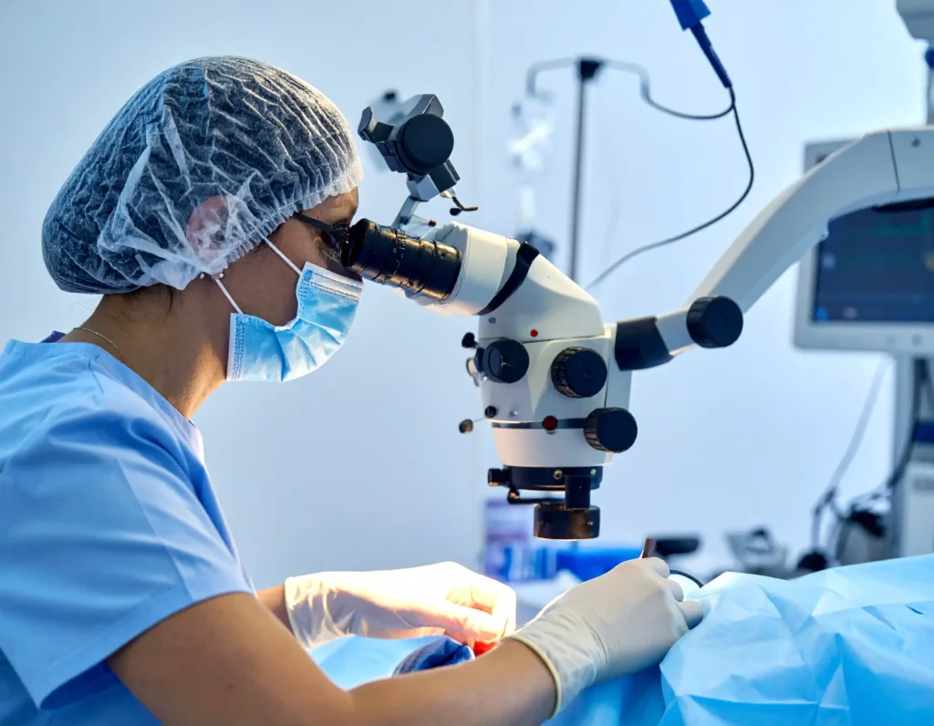
Caries treatment under a microscope is a micro-surgical procedure in a field that is maximally illuminated and magnified, which allows for more precise detection and selective removal of carious tissues, while protecting healthy structures and the pulp thanks to better control of the final cavity preparation "end-point" and the marginal seal of the restoration. In practice, modern protocols of selective dentin removal are combined with adhesive composite restoration, and magnification facilitates the assessment of cracks, secondary caries at the margins, and material adaptation, which promotes a minimally invasive approach and the maintenance of pulp vitality.
What it involves
The operating microscope provides 3–30× magnification and coaxial light, which improves the visibility of fine structures, facilitating diagnostics and micro-manipulations in cavity preparation and adhesive composite restoration. In conservative treatment under a microscope, the minimally invasive concept dominates: cleaning the periphery of the cavity to hard dentin for a tight marginal zone and selective leaving of leathery/soft dentin over the pulp depending on the depth. The microscope also facilitates control of the matrix, layering, marginal fit, and finishing, reducing the risk of micro-leakage and iatrogenic damage.
Key clinical benefits
Greater precision and tissue preservation thanks to better visibility, which limits over-preparation and reduces the risk of pulp exposure in deep cavities.
Better assessment and finishing of margins, composite adaptation, and detection of cracks and secondary caries, which supports long-term marginal integrity.
Minimally invasive selective preparation techniques are associated with higher clinical efficacy and a lower frequency of pulp exposures compared to complete caries removal.
Ergonomics and documentation: improved working posture and the possibility of photo/video documentation to facilitate patient education and communication.
When it is particularly beneficial
Deep cavities with a risk of pulp exposure, where selective removal to firm or soft dentin is preferred to maintain vitality.
Margin control in adhesive reconstructions and verification of secondary caries and enamel/dentin micro-cracks.
Situations requiring perfect adaptation and smaller marginal gaps, e.g., preparations for indirect restorations and CAD/CAM.
Increased magnification narrows the field of view and depth of field. Research on caries diagnostics suggests that extremely high magnifications do not necessarily improve diagnostic accuracy and may even lead to over-diagnosis of lesions; therefore, the choice of magnification should be purposeful. Using magnification also improves the clinician's ergonomics and posture.
In restorations, microscope-aided work is associated with better adaptation control and can reduce marginal gaps in preparations for CAD/CAM work, as confirmed by micro-CT analyses and quality studies of posterior fillings.
Microscopic lighting with a yellow filter prevents premature polymerization of light-cured materials and improves visual comfort, while green filters can facilitate tissue differentiation during surgical procedures.
Rycerska 4 Medical Center in Bytom is a modern facility trusted by over 8,000 patients, offering a wide range of services, an experienced team of doctors and therapists, convenient online registration, telephone contact and via WhatsApp, as well as support in treatment financing and the possibility of purchasing gift vouchers for treatments at the clinic.
Explore the details of our offer in the following areas:
- comprehensive conservative dentistry
- comprehensive dental prosthetics
- dental surgery
- modern implantology
- comprehensive treatment under a microscope
- endodontics
- comprehensive dental prophylaxis
- professional hygienization
- comprehensive RTG and CBCT (tomography) diagnostics
- dental physiotherapy
- aesthetic medicine
- aesthetic facial physiotherapy
- general physiotherapy, pain therapy
- massages
| Procedure name | Price |
|---|---|
| Infiltration and block anesthesia | 50 PLN |
| Chemical-cured filling | 200 – 250 PLN |
| Light-cured composite filling, 1-surface – small | 250 PLN |
| Light-cured composite filling, 2-surface – medium | 300 PLN |
| Light-cured composite filling, 3-surface – large | 350 PLN |
| 4-surface light-cured filling – reconstruction of a severely damaged tooth | 400 zł |
| Temporary filling – medicinal dressing | 150 zł |
| Splinting of teeth (per space) | 200 zł |
| Biological pulp treatment (MTA/Biodentine) with light-cured filling | 300 – 600 zł |
| Each subsequent glass fiber | 150 zł |
| ICON (treatment of early caries lesions/removal of discolorations) per 1 tooth | from 250 zł |
Painless treatment
Aesthetic and natural restorations
Precise diagnostics
Comprehensive offer

See reviews
Looking for an ideafor a gift?


Select voucher type

Complete the details and pay for the order

Collect the voucher online or on-site