Microscopic treatment
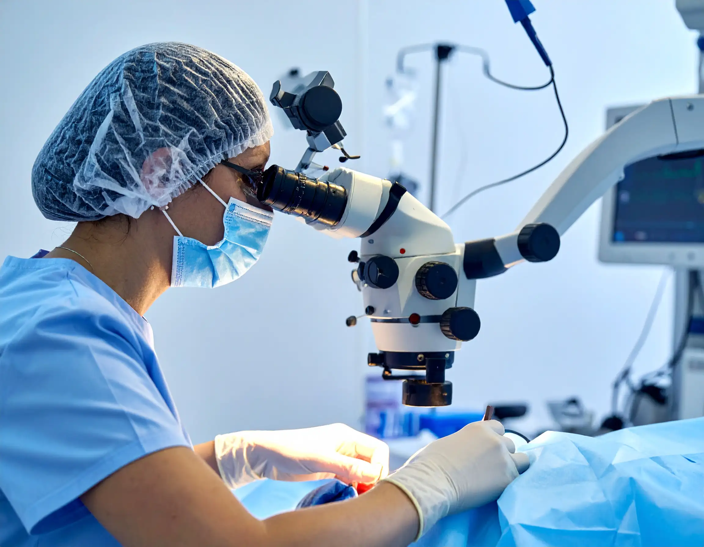
Microscopic dental treatment (microscopic endodontics) involves performing dental procedures – most often root canal treatment – using an operating microscope. This allows for several-fold magnification of the operating field (up to 25 times), enabling the doctor to more precisely locate, clean, and disinfect the root canals and subsequently fill them.
Benefits of using a mikroskop
Microscopic dental treatment (microscopic endodontics) involves performing dental procedures – most often root canal treatment – using an operating microscope. This allows for several-fold magnification of the operating field (up to 25 times), enabling the doctor to more precisely locate, clean, and disinfect the root canals and subsequently fill them.
The primary benefits of treatment under a microscope are greater precision of the procedure, minimisation of the risk of complications and reinfection (it often allows saving a tooth that would have to be removed using classic methods), the ability to identify details invisible to the naked eye (e.g., micro-cracks, additional canals or branches affecting the effectiveness of therapy), increased safety and comfort (the procedure is painless, performed in a well-lit and magnified area), shortening of treatment time and the reduction of necessary repeat visits (treatment is sometimes even performed in a single visit),
Centrum Medyczne Rycerska 4 in Bytom is a modern facility trusted by over 8,000 patients, offering a wide range of services, an experienced team of doctors and therapists, convenient online registration, phone and WhatsApp contact, as well as support in treatment financing and the option to purchase gift vouchers for treatments at the facility.
Find out more about our offer in the following areas:
- comprehensive conservative dentistry
- comprehensive dental prosthetics
- dental surgery
- modern implantology
- comprehensive treatment under a microscope
- endodontics
- comprehensive dental prophylaxis
- professional hygienization
- comprehensive RTG and CBCT (tomography) diagnostics
- dental physiotherapy
- aesthetic medicine
- aesthetic facial physiotherapy
- general physiotherapy, pain therapy
- massages
Painless treatment
Aesthetic and natural restorations
Precise diagnostics
Comprehensive offer
Price list
| Procedure name | Price |
|---|---|
| Root canal treatment (endodontics) of tooth 1 – 3 (incisors, canines – including RTG and anesthesia) | 700 – 1000 zł |
| Root canal treatment (endodontics) of tooth 4 – 5 (premolars – including RTG and anesthesia) | 900 – 1200 zł |
| Root canal treatment (endodontics) of tooth 6 – 8 (molars – including RTG and anesthesia) | 1100 – 1500 zł |
| Re-treatment (root canal) of tooth 1 – 3 (incisors, canines – including RTG and anesthesia) | 900 - 1300 zł |
| Re-treatment (root canal) of tooth 4 – 5 (premolars – including RTG and anesthesia) | 1100 - 1500 zł |
| Re-treatment (root canal) of tooth 6 – 8 (molars – including RTG and anesthesia) | 1300 - 1700 zł |
| Removal of a broken instrument from the tooth canal (during the procedure) | 300 – 700 zł |
| Closure of perforation with biological material | 200 – 300 zł |
| Glass fiber post (without tooth reconstruction) | 300 PLN |
| Tooth core buildup before root canal treatment (endodontic) | 250 - 450 zł |
| Reconstruction of a severely damaged tooth after root canal treatment (endodontic) | 400 zł |
| Reconstruction of a severely damaged tooth after root canal treatment (endodontic) using glass fiber | 600 zł |
When to use a mikroskop
mikroskop treatment is superior to traditional treatment, especially in cases of complex anatomy or high risk of complications, as it increases precision, reduces invasiveness, and improves the predictability of clinical outcomes.
In uncomplicated class I–II cavities, simple single-rooted canals, and routine check-ups, the safety and efficacy of the method without a mikroskop may be sufficient, although magnification can still be diagnostically helpful. The decision should be based on the difficulty of the case, the risk of complications, and the need to preserve as much tissue as possible.
In cases of complex anatomy and difficult canals, the mikroskop wins, especially when there are additional, obliterated, or narrow canals, saddle-shaped chambers, curved roots, or branching, where missing structures is common without magnification. Magnification and shadowless lighting facilitate the localization of canal orifices, orientation in anomalies, and minimization of healthy tissue loss.
During reendo, after traditional treatment failure, the mikroskop improves the chances of removing gutaperka, sealing materials, posts, and broken instruments, as well as closing perforations and controlling bleeding. This reduces the risk of subsequent failures due to more thorough disinfection and tighter filling of the canal system.
Detection of micro-cracks, marginal gaps, and micro-leakage around fillings, crowns, or onlays is more effective under magnification, allowing for more conservative, precise preparation and longer durability of restorations. Better visibility limits excessive preparation and supports minimally invasive adhesive techniques.
Endodontyczna and periodontologiczne mikrochirurgia – in procedures of apicoectomy, removal of periapical granulomas/cysts, and periodontal mikrochirurgia, the mikroskop increases control over the incision, hemostasis, and suturing, which shortens healing and improves aesthetic outcomes. Precise magnification is key when working in the apical area and during soft tissue micro-grafts.
Conservative and aesthetic precision during caries removal at the dentin-enamel junction, preparation for veneers/crowns, and marginal fit. This reduces the risk of leaving infected tissues and supports the longevity of the work. Magnification allows for preserving more healthy structure, which is an advantage over the traditional approach without optics.
Less invasiveness and better predictability – thanks to better insight, procedures are shorter, more tissue-sparing, and carry a lower risk of perforation, missed canals, or instrument breakage compared to methods without a mikroskop. This translates to fewer re-interventions and higher long-term clinical efficacy and patient comfort.
Typical practical indications:
-
- Reendo, perforations, removal of broken instruments, atypical anatomy, strongly curved canals.
- Symptoms suggesting a crown/root fracture or recurring ailments despite correct RVG/CBCT images.
- Periapical mikrochirurgia
- Periodontologiczne procedures requiring precise suturing and atraumatic technique.
Centrum Medyczne Rycerska 4 in Bytom is a modern facility trusted by over 8,000 patients, offering a wide range of services, an experienced team of doctors and therapists, convenient online registration, phone and WhatsApp contact, as well as support in treatment financing and the option to purchase gift vouchers for treatments at the facility.
Find out more about our offer in the following areas:
- comprehensive conservative dentistry
- comprehensive dental prosthetics
- dental surgery
- modern implantology
- comprehensive treatment under a microscope
- endodontics
- comprehensive dental prophylaxis
- professional hygienization
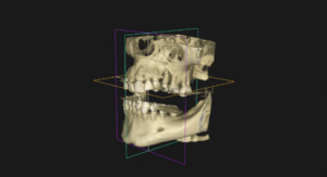
- comprehensive RTG and CBCT (tomography) diagnostics
- dental physiotherapy
- aesthetic medicine
- aesthetic facial physiotherapy
- general physiotherapy, pain therapy
- massages
Painless treatment
Aesthetic and natural restorations
Precise diagnostics
Comprehensive offer
Price list
| Procedure name | Price |
|---|---|
| Root canal treatment (endodontics) of tooth 1 – 3 (incisors, canines – including RTG and anesthesia) | 700 – 1000 zł |
| Root canal treatment (endodontics) of tooth 4 – 5 (premolars – including RTG and anesthesia) | 900 – 1200 zł |
| Root canal treatment (endodontics) of tooth 6 – 8 (molars – including RTG and anesthesia) | 1100 – 1500 zł |
| Re-treatment (root canal) of tooth 1 – 3 (incisors, canines – including RTG and anesthesia) | 900 - 1300 zł |
| Re-treatment (root canal) of tooth 4 – 5 (premolars – including RTG and anesthesia) | 1100 - 1500 zł |
| Re-treatment (root canal) of tooth 6 – 8 (molars – including RTG and anesthesia) | 1300 - 1700 zł |
| Removal of a broken instrument from the tooth canal (during the procedure) | 300 – 700 zł |
| Closure of perforation with biological material | 200 – 300 zł |
| Glass fiber post (without tooth reconstruction) | 300 PLN |
| Tooth core buildup before root canal treatment (endodontic) | 250 - 450 zł |
| Reconstruction of a severely damaged tooth after root canal treatment (endodontic) | 400 zł |
| Reconstruction of a severely damaged tooth after root canal treatment (endodontic) using glass fiber | 600 zł |
Differences in efficacy
In primary root canal treatment, efficacy is high in both approaches, but the mikroskop increases the success rate, especially in molars and complex cases; in reendo and mikrochirurgia, the advantage of the mikroskop is clear and confirmed by clinical studies.
Primary treatment (RCT)
Contemporary root canal treatment has high overall survival/success rates; meta-analyses and reviews report survival >90–97% in the long term with proper restoration, although methodologies differ in definitions of clinical vs radiological success.
In comparisons of mikroskop vs without a mikroskop, studies show a higher percentage of positive outcomes using a mikroskop, especially in posterior teeth; in one cohort for molars, the mikroskop increased the success rate 2.9–3.2 times compared to the traditional method under strict and liberal criteria.
Retreatment (reendo)
The efficacy of reendo is lower than primary treatment, but magnification improves the prognosis due to easier localization of missed canals, removal of posts/material, and perforation control; clinical reports indicate higher success rates for reendo with a mikroskop vs without.
In difficult cases (obliterated canals, broken instruments), mikroskop-assisted techniques effectively resolve 70+% of cases, e.g., 74% for calcified canals and 72% for separated instruments in case series analyses.
Surgical mikroendodoncja
Classical periapical surgery has an efficacy of 44–75% in older studies, while endodontyczna microsurgery (mikroskop, micro-instrumentarium, retrograde US filling) reaches 57–97% depending on criteria and observation time.
Clinical series descriptions also indicate faster and more predictable healing with the mikrochirurgia technique, which is attributed precisely to the precision of the optics and technique, rather than just the materials.
What determines the differences? Visibility of additional canals and anatomical anomalies increases the completeness of the preparation and the tightness of the filling, which directly raises efficacy and lowers the risk of recurrent periapical lesions.
The impact of the mikroskop is greatest in molars, cases with large periapical lesions, in reendo, and in surgery; in simple single-canal incisors, the differences may be clinically smaller with a good protocol, but magnification still reduces the risk of errors.
Centrum Medyczne Rycerska 4 in Bytom is a modern facility trusted by over 8,000 patients, offering a wide range of services, an experienced team of doctors and therapists, convenient online registration, phone and WhatsApp contact, as well as support in treatment financing and the option to purchase gift vouchers for treatments at the facility.
Find out more about our offer in the following areas:
- comprehensive conservative dentistry
- comprehensive dental prosthetics
- dental surgery
- modern implantology
- comprehensive treatment under a microscope
- endodontics
- comprehensive dental prophylaxis
- professional hygienization
- comprehensive RTG and CBCT (tomography) diagnostics
- dental physiotherapy
- aesthetic medicine
- aesthetic facial physiotherapy
- general physiotherapy, pain therapy
- massages
Painless treatment
Aesthetic and natural restorations
Precise diagnostics
Comprehensive offer
Price list
| Procedure name | Price |
|---|---|
| Root canal treatment (endodontics) of tooth 1 – 3 (incisors, canines – including RTG and anesthesia) | 700 – 1000 zł |
| Root canal treatment (endodontics) of tooth 4 – 5 (premolars – including RTG and anesthesia) | 900 – 1200 zł |
| Root canal treatment (endodontics) of tooth 6 – 8 (molars – including RTG and anesthesia) | 1100 – 1500 zł |
| Re-treatment (root canal) of tooth 1 – 3 (incisors, canines – including RTG and anesthesia) | 900 - 1300 zł |
| Re-treatment (root canal) of tooth 4 – 5 (premolars – including RTG and anesthesia) | 1100 - 1500 zł |
| Re-treatment (root canal) of tooth 6 – 8 (molars – including RTG and anesthesia) | 1300 - 1700 zł |
| Removal of a broken instrument from the tooth canal (during the procedure) | 300 – 700 zł |
| Closure of perforation with biological material | 200 – 300 zł |
| Glass fiber post (without tooth reconstruction) | 300 PLN |
| Tooth core buildup before root canal treatment (endodontic) | 250 - 450 zł |
| Reconstruction of a severely damaged tooth after root canal treatment (endodontic) | 400 zł |
| Reconstruction of a severely damaged tooth after root canal treatment (endodontic) using glass fiber | 600 zł |
Endodoncja under a mikroskop
Root canal treatment (also known as endodoncja) is a procedure that saves the tooth from loss (extraction), consisting of removing infected or dead pulp, thorough preparation (cleaning), and disinfection of root canals, as well as their tight filling and reconstruction of the tooth crown.
When it is necessary
Root canal (endodontyczne) treatment is performed when decay, trauma, or a crack leads to inflammation or necrosis of the pulp, manifesting as pain, sensitivity, swelling, or the presence of periapical lesions in imaging studies. Indications also include failures of earlier therapy and ailments despite correct fillings and crowns.
Root canal treatment under a mikroskop (microscopic endodoncja) allows for several-fold magnification of the operative field (up to 25 times), enabling the doctor to more precisely locate, clean, and disinfect the tooth canals and subsequently fill them.
The basic benefits of treatment under a mikroskop are:
-
- greater precision of the procedure,
- minimization of the risk of complications and re-infection (often allowing the tooth to be saved, which would have had to be extracted using classical methods),
- possibility of identifying details invisible to the naked eye (e.g., micro-cracks, additional canals, or branches affecting the therapy's success)
- increased safety and comfort (the procedure is painless, performed in a well-lit and magnified area),
- shortening of the treatment time and the need for repeat visits (treatment is sometimes performed even in a single visit),
Step-by-step process
-
- Comprehensive diagnostics: interview, clinical examination, RVG/CBCT image to assess anatomy, working length, and periapical lesions.
- Anesthesia and koferdam isolation: ensuring comfort and an aseptic treatment field.
- Opening the chamber and removing the pulp: gaining access to the canals and eliminating infected tissues, preferably under mikroskop control.
- Preparation and disinfection: mechanical-chemical shaping of canals with manual/rotary instruments and irrigation with antiseptic agents.
- Filling: tight obturation (filling) with gutta-percha and sealer, radiological control.
- Restoration: from filling to post and crown, depending on tissue loss.
Time and number of visits
A simple case can be completed in a single visit, while complex cases involving complicated anatomy, root canal re-treatment, or acute symptoms often require 2–3 appointments with intermediate treatment (dressings).
Effectiveness and prognosis
Properly performed root canal treatment achieves high success rates with appropriate restoration and marginal seal, whereas more difficult cases (large lesions, posterior teeth, reendo) have a lower prognosis without magnification and thorough disinfection. The use of a microscope and CBCT increases predictability through better canal localization and more precise quality control.
Possible complications
Risks include:
-
- missed or hard-to-reach canal,
- perforation,
- instrument fracture,
- insufficient disinfection,
- obturation leakage
- root fractures.
This may result in the persistence of lesions or the need for re-treatment or possibly microsurgery. In case of failure, re-treatment, apicoectomy, or ultimately extraction and implantation or another prosthetic replacement is possible.
What determines success
-
- Comprehensive diagnostic imaging and proper identification of canal anatomy.
- Proper asepsis and rubber dam isolation, as well as effective irrigation.
- Complete and duly diligent preparation of all canals and tight obturation (filling of the canals).
- Post-endodontic restoration ensuring strength and marginal seal, ideally as soon as possible after treatment.
Centrum Medyczne Rycerska 4 in Bytom is a modern facility trusted by over 8,000 patients, offering a wide range of services, an experienced team of doctors and therapists, convenient online registration, phone and WhatsApp contact, as well as support in treatment financing and the option to purchase gift vouchers for treatments at the facility.
Find out more about our offer in the following areas:
- comprehensive conservative dentistry
- comprehensive dental prosthetics
- dental surgery
- modern implantology
- comprehensive treatment under a microscope
- endodontics
- comprehensive dental prophylaxis
- professional hygienization
- comprehensive RTG and CBCT (tomography) diagnostics
- dental physiotherapy
- aesthetic medicine
- aesthetic facial physiotherapy
- general physiotherapy, pain therapy
- massages
Painless treatment
Aesthetic and natural restorations
Precise diagnostics
Comprehensive offer
Price list
| Procedure name | Price |
|---|---|
| Root canal treatment (endodontics) of tooth 1 – 3 (incisors, canines – including RTG and anesthesia) | 700 – 1000 zł |
| Root canal treatment (endodontics) of tooth 4 – 5 (premolars – including RTG and anesthesia) | 900 – 1200 zł |
| Root canal treatment (endodontics) of tooth 6 – 8 (molars – including RTG and anesthesia) | 1100 – 1500 zł |
| Re-treatment (root canal) of tooth 1 – 3 (incisors, canines – including RTG and anesthesia) | 900 - 1300 zł |
| Re-treatment (root canal) of tooth 4 – 5 (premolars – including RTG and anesthesia) | 1100 - 1500 zł |
| Re-treatment (root canal) of tooth 6 – 8 (molars – including RTG and anesthesia) | 1300 - 1700 zł |
| Removal of a broken instrument from the tooth canal (during the procedure) | 300 – 700 zł |
| Closure of perforation with biological material | 200 – 300 zł |
| Glass fiber post (without tooth reconstruction) | 300 PLN |
| Tooth core buildup before root canal treatment (endodontic) | 250 - 450 zł |
| Reconstruction of a severely damaged tooth after root canal treatment (endodontic) | 400 zł |
| Reconstruction of a severely damaged tooth after root canal treatment (endodontic) using glass fiber | 600 zł |
Caries treatment under a microscope
Caries treatment under a microscope is a micro-surgical procedure in a field that is maximally illuminated and magnified, which allows for more precise detection and selective removal of carious tissues, while protecting healthy structures and the pulp thanks to better control of the final cavity preparation "end-point" and the marginal seal of the restoration. In practice, modern protocols of selective dentin removal are combined with adhesive composite restoration, and magnification facilitates the assessment of cracks, secondary caries at the margins, and material adaptation, which promotes a minimally invasive approach and the maintenance of pulp vitality.
What it involves
The operating microscope provides 3–30× magnification and coaxial light, which improves the visibility of fine structures, facilitating diagnostics and micro-manipulations in cavity preparation and adhesive composite restoration. In conservative treatment under a microscope, the minimally invasive concept dominates: cleaning the periphery of the cavity to hard dentin for a tight marginal zone and selective leaving of leathery/soft dentin over the pulp depending on the depth. The microscope also facilitates control of the matrix, layering, marginal fit, and finishing, reducing the risk of micro-leakage and iatrogenic damage.
Key clinical benefits
Greater precision and tissue preservation thanks to better visibility, which limits excessive preparation and reduces the risk of pulp exposure in deep cavities.
Better assessment and processing of margins, composite adaptation, and detection of cracks and secondary caries, which supports durable marginal seals.
Minimally invasive selective preparation techniques are associated with higher clinical effectiveness and a lower incidence of pulp exposure compared to complete caries removal.
Ergonomics and documentation: improvement of the working position and the possibility of photo/video documentation facilitating education and communication with the patient.
When it is particularly beneficial
Deep cavities with a risk of pulp exposure, where selection to firm or soft dentin is preferred to maintain vitality.
Control of margins in adhesive reconstructions and verification of secondary caries and enamel/dentin micro-cracks.
Situations requiring perfect adaptation and smaller marginal gaps, e.g., preparations for indirect restorations and CAD/CAM.
Increasing magnification narrows the field of vision and depth of field. Research on caries diagnostics suggests that extremely high magnifications do not necessarily improve diagnostic accuracy and may even promote overdiagnosis of lesions, so the choice of magnification should be purposeful. The use of magnification also improves ergonomics and the doctor's posture.
In restorations, microscope support is associated with better control of adaptation and can reduce marginal gaps in preparations for CAD/CAM work, as confirmed by micro-CT analyses and studies of posterior filling quality.
Microscopic lighting with a yellow filter prevents premature polymerization of light-cured materials and improves visual comfort, while green filters can facilitate tissue differentiation during surgical procedures.
Centrum Medyczne Rycerska 4 in Bytom is a modern facility trusted by over 8,000 patients, offering a wide range of services, an experienced team of doctors and therapists, convenient online registration, phone and WhatsApp contact, as well as support in treatment financing and the option to purchase gift vouchers for treatments at the facility.
Find out more about our offer in the following areas:
- comprehensive conservative dentistry
- comprehensive dental prosthetics
- dental surgery
- modern implantology
- comprehensive treatment under a microscope
- endodontics
- comprehensive dental prophylaxis
- professional hygienization
- comprehensive RTG and CBCT (tomography) diagnostics
- dental physiotherapy
- aesthetic medicine
- aesthetic facial physiotherapy
- general physiotherapy, pain therapy
- massages
Painless treatment
Aesthetic and natural restorations
Precise diagnostics
Comprehensive offer
Price list
| Procedure name | Price |
|---|---|
| Infiltration and block anesthesia | 50 PLN |
| Chemical-cured filling | 200 – 250 PLN |
| Light-cured composite filling, 1-surface – small | 250 PLN |
| Light-cured composite filling, 2-surface – medium | 300 PLN |
| Light-cured composite filling, 3-surface – large | 350 PLN |
| 4-surface light-cured filling – reconstruction of a severely damaged tooth | 400 zł |
| Temporary filling – medicinal dressing | 150 zł |
| Splinting of teeth (per space) | 200 zł |
| Biological pulp treatment (MTA/Biodentine) with light-cured filling | 300 – 600 zł |
| Each subsequent glass fiber | 150 zł |
| ICON (treatment of early caries lesions/removal of discolorations) per 1 tooth | from 250 zł |