RTG and CBCT diagnostics
Dental RTG, or dental radiography, is one of the most important diagnostic tests in dentistry. It enables detailed imaging of teeth, roots, jaw and mandible bones, and surrounding tissues. This allows the doctor to precisely assess the condition of the teeth, detect changes invisible to the naked eye, and plan effective treatment.
RTG diagnostics, alongside a thorough medical interview, precise clinical examination, and analysis of subjective and objective symptoms reported by the patient – constitutes one of the foundations of correct diagnosis and effective treatment. Today, meeting our patients' needs and constantly improving the quality of our services, we are introducing CBCT technique imaging to the Rycerska 4 Medical Center.
Learn more
RTG diagnostics is one of the foundations of an accurate diagnosis. Patient awareness regarding the immense diagnostic value provided by modern imaging techniques is still insufficient, and doctors of various specialties, especially dentists, are frequently questioned about the necessity of expanding diagnostics by performing RTG scans.
expand to learn more about RTG diagnostics
Many clinical conditions in dental practice are impossible to diagnose without correctly performed RTG images, and only proper diagnostic imaging enables their detection, which subsequently implies specific therapeutic procedures.
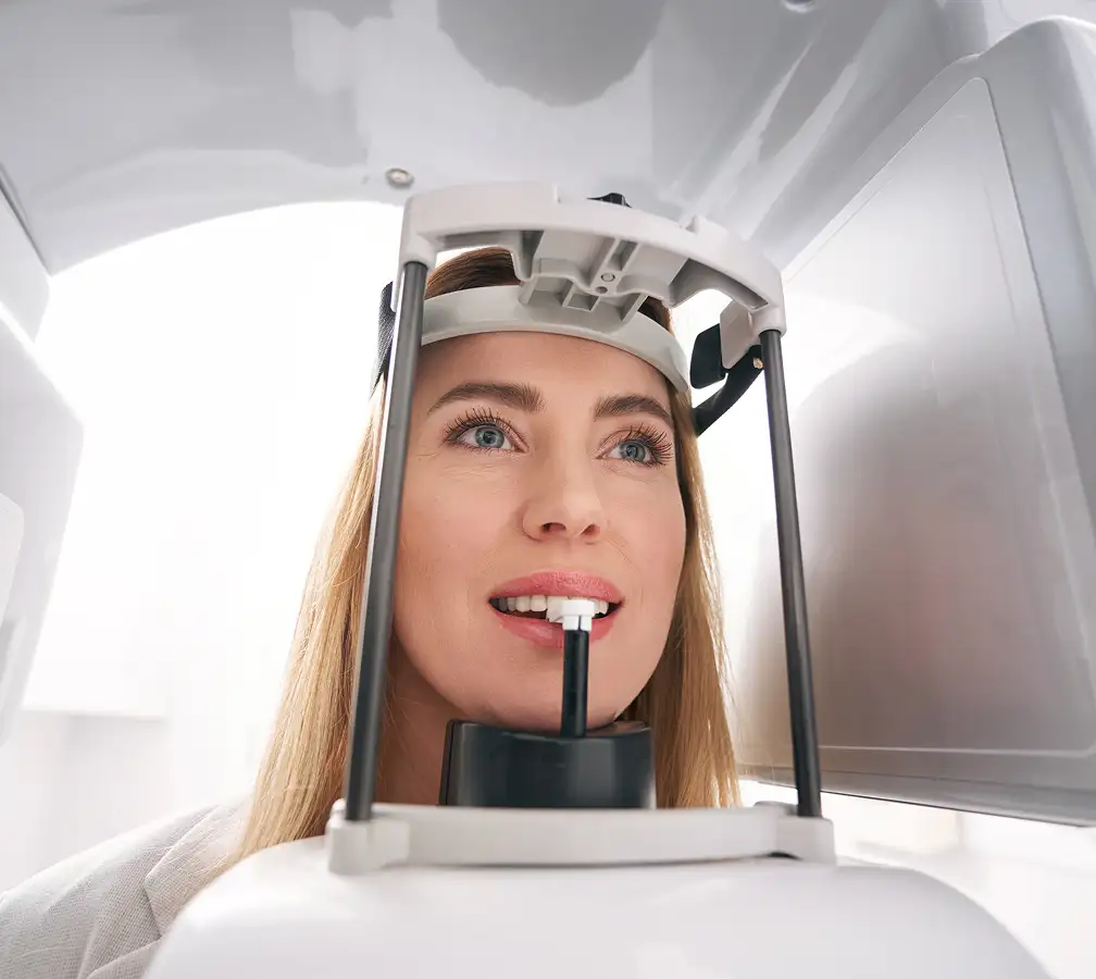
Techniques using X-ray radiation find particular application in dental practice, ranging from 2D images, such as periapical RTG, bitewing RTG, and orthopantomographic RTG (so-called panorama), to the significantly more accurate and everyday-implemented 3D imaging. 3D imaging using computed tomography techniques is currently one of the main standards of precision dentistry. Sections available through the use of the CBCT (Cone Beam Computed Tomography) technique, i.e., the cone-beam technique, allow for a precise analysis of the diagnosed area while reducing the radiation dose compared to a classic head CT, which is primarily why they are widely used in all fields of dentistry today.
Below you can find basic information about the most common types of RTG performed at our facility.
Periapical RTG (point, dental):
expand to see more information…
- performed using a radiation detector placed inside the patient's oral cavity adjacent to the part of the dental arch to be imaged
- can be performed using Cieszynski's isometry technique (bisecting angle) or the paralleling technique
- allow for imaging of a small field; most commonly used for the diagnostics of a single tooth.
Bitewing RTG:
expand to learn more about bitewing radiographs …
- allows for simultaneous visualization of the crowns of the upper and lower teeth along with the marginal periodontium
- particularly valuable for the diagnosis of interproximal caries, secondary caries under fillings, and the assessment of the subgingival margins of fillings
- RTG films have special “wings” (bite tabs) used to hold the film between the upper and lower teeth.
Occlusal RTG:
check for more information on occlusal radiographs…
- uses a radiation detector placed in the oral cavity in the occlusal plane
- have very specific indications for their performance, such as:
In the case of maxilla diagnostics:
- Assessment of the position of impacted teeth (buccal/palatal position), supernumerary teeth, supplemental teeth, and odontomas.
- Determining the extent of lesions, e.g., cysts, tumors.
- Diagnostics of cleft palate.
- Assessment of the periapical area.
- Diagnostics of tooth and alveolar process fractures.
- Detection and assessment of the position of tooth roots displaced into the maxillary sinus lumen.
- Auxiliary assessment of tooth root positions.
In the case of mandible diagnostics:
- Assessment and location of salivary stones in the salivary gland ducts.
- Assessment of the position of impacted teeth (buccal/lingual position); especially third molars.
- Determining the extent of lesions, e.g., cysts, tumors.
- Assessment of the periapical area.
- Diagnostics of tooth and alveolar process fractures.
Orthopantomographic RTG (so-called panorama, OPG):
expand to learn more about panoramic radiographs …
- it is a radiograph obtained through the reciprocal movement of the cassette and the X-ray tube around the examined object along an elliptical path
- it is a layered radiograph in which the teeth, maxillary alveolar process, and the alveolar part of the mandible are visualized simultaneously, as well as the craniofacial bones including the maxillary sinuses and temporomandibular joints
- is not a suitable tool for diagnosing the anterior section of the dental arches, i.e., from the canine to the canine, due to image blurring
- allows for the detection of many changes such as bone rarefaction and condensation, which, however, usually require detailed diagnostics within 3D imaging.
Cone-Beam Tomography (cone-beam tomography, CBCT):
check for more information on CBCT imaging…
- this is a tomography performed using a cone-shaped X-ray beam emitted by an X-ray tube rotating around the patient's head
- short exposure time (10-40 seconds) allows for a reduction in the ionizing radiation dose (compared to classic head CT) and reduces the number of artifacts
- is widely used in all fields of dentistry, including endodontics, dental surgery with implantology, and even daily restorative dentistry.
Applications in endodontics:
- Localization of hidden, additional, and atypically located or coursing root canals.
- Determining the actual number of root canals and their course.
- Detection of early inflammatory processes in periapical tissues, invisible in 2D imaging.
- Diagnosis of root fractures and their course (significant for qualifying for endodontic treatment).
- Determining the extent of lesions and – by comparing images over time – precise assessment of the healing progress (or lack thereof).
- Diagnostics of complications and a more precise assessment of prognosis.
Applications in dental surgery and implantology:
- Precise determination of the position of tooth roots and impacted teeth (as well as additional, supernumerary teeth, etc.) and their position relative to neighboring teeth and strategic anatomical structures, such as the mandibular nerve canal, maxillary sinus, etc.
- Assessment of bone quantity and qualitative suitability for implantation
- Assessment of post-treatment healing progress (or lack thereof)
- Assessment of the extent of inflammatory and neoplastic changes
- Assessment of complications and a more precise evaluation of prognosis
Applications in conservative dentistry:
- Assessment of the extent of the carious process.
- Detection of early inflammatory processes in periapical tissues, invisible in 2D imaging
Note !!! RTG does not detect inflammatory changes occurring in the dental pulp, but only their causes, such as a very deep carious lesion, or the consequences of a spreading inflammatory process.
At Rycerska 4 Medical Center, we require a referral for RTG examinations, which precisely specifies the scope of diagnostics. A referral template can be downloaded here.
RTG examinations do not require special preparation from the Patient; immediately before the procedure, you should remove any items that could potentially affect the quality of the examination (e.g., jewelry, hearing aids, removable prosthetic restorations)
Dental RTG examinations are safe procedures, however – like any procedure using ionizing radiation – they require individual patient qualification. Contraindications primarily concern pregnant women, which is related to the possibility of a negative impact of radiation on the developing fetus. RTG diagnostics in pregnant women are performed only in situations where the benefits outweigh the potential risk, and the final decision to conduct the RTG examination is made by the doctor.
Patients from Bytom and the entire Silesia region choose our clinic because they know they can count on safety, empathy, and a speedy recovery.